Search from website

Mouse mAb hGDNF (clone 3C1)

Mouse mAb hGDNF (clone 3C1)  
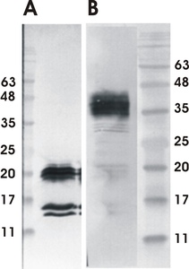
Figure 1. Western Blot testing of GDNF monoclonal antibody 3C1. Analysis was performed with antiGDNF monoclonal antibody 3C1 HRP conjugate. 15 µl of CHOEBNALT85 GDNF producing cell culture supernatant was loaded per lane. Analysis was performed in reducing (A) and non-reducing (B) conditions
A
Mouse mAb hGDNF (clone 3C1)
 
B
Mouse mAb hGDNF (clone 3C1)
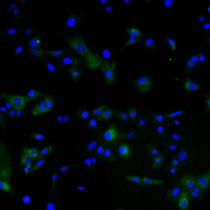
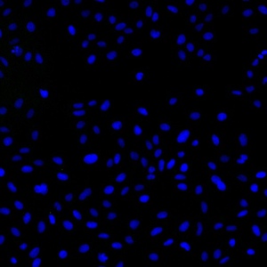
Figure 2.Immunofluorescence detection of human GDNF expressed in U2OS cells by anti-GDNF monoclonal antibody 3C1. Anti-GDNF antibody 3C1 concentration in IF experiment was 10 µg/ml. Goat ant-mouse AlexaFluor488 was used as secondary antibody. For nuclear staining DAPI was used. ArrayScan VTI platform (Thermo Scientific) was used for image acquisition (10x objective). Composite picture was generated using pseudocolors green for GDNF specific signal and blue for nuclei. A. GDNF-expressing U2OS cells; B. Negative control (non-transfected U2OS cells)
A
Mouse mAb hGDNF (clone 3C1)
B
Mouse mAb hGDNF (clone 3C1)
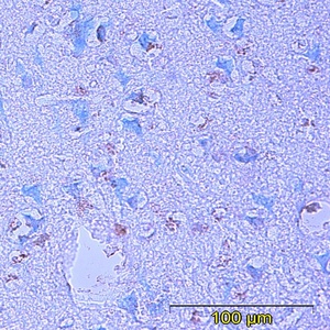
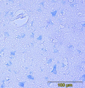
Figure 3. Immunohistochemistry testing of anti-GDNF monoclonal antibody 3C1. Analysis was performed using formalin-fixed paraffin-embedded human cerebral cortex tissue sections from Alzheimer’s disease patients. Tissue sections were boiled with sodium citrate buffer (pH 6) for antigen retrieval. Incubation with primary antibody at 5 µg/ml was performed overnight at 4°C. DAKO EnVisionTM Detection System, Peroxidase/DAB was used for visualization. Sections were counterstained with toluidine blue and mounted with Eukitt mounting medium. A. GDNF staining by monoclonal antibody 3C1; B.Negative staining without primary antibody

Select volume:

Quantity:

€ 2307

Catalogue #

315-100

Name

Mouse mAb hGDNF (clone 3C1)

Target

Human GDNF

Target Description

Recombinant human GDNF protein produced using E. coli expression system

Alternative Names

Astrocyte-derived trophic factor (ATF)

Uniprot ID

P39905

Clonality

Mouse monoclonal

Clone

3C1

Class

mIgG1

Reactivity

Human GDNF

Application

ELISA, WB, IF, IHC

Protocol

Monoclonal antibody working amount has to be established practically for each particular antigen and assay format

ELISA:

50-100 ng/ml

WB:

2-5 µg/ml

IF:

2-10 µg/ml

IHC:

5 µg/ml

Purification

Protein G purification

Buffer

PBS pH 7.4, with 0.1% sodium azide

Shipping

This product is shipped in non-frozen liquid form in ambient conditions

Storage

Store at –20…-70°C upon receipt. Divide antibody into aliquots prior usage. Avoid multiple freeze-thaw cycles

Background

GDNF is a neurotrophic factor that enhances survival and morphological differentiation of dopaminergic neurons and increases their high-affinity dopamine uptake. Ligand for the GFR-alpha-3-RET receptor complex but can also activate the GFR-alpha-1-RET receptor complex

This product is for research use only

TECHNICAL ASSISTANCE

Please refer any technical questions to

technical.support@icosagen.com

Our locations

United States Office

77 Geary St. 5th Floor
San Francisco, CA 94108
+1 415 548 6625

European Office

Eerika tee 1
Õssu, Kambja vald
Tartu maakond 61713
Estonia
+372 737 7070
Shopping Cart