
Search from website
Search from website
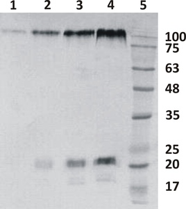
Figure 1. Western-Blot analysis of Ebolavirus GP protein using Rabbit polyclonal antibody to Zaire Ebolavirus PG protein, A2-100-100. Different amounts of recombinant GP protein is loaded per lane. Analysis is performed in reducing conditions. Antibody is detecting both, GP1 and GP2 proteins. Lane 1. 10 ng; Lane 2. 50 ng; Lane 3. 100 ng; Lane 4. 200 ng; Lane 5. Prestained Protein Ladder, Naxo 8003. Primary antibody dilution 0.2 µg/ml was used.
Catalogue #
A2-100-100
Name
Rabbit pAb to Zaire Ebolavirus GP
Target
Ebolavirus GP protein
Target Description
Envelope glycoprotein processed to GP1 and GP2. Transmembrane and cytoplasmatic domains are removed. Immunogen contains C-terminal linker and hexahistidine tag sequence (GSGHHHHHH). Expressed by 293-based cell line (expressed by QMCF Technology)
Alternative Names
Virion spike glycoprotein, GP protein
Uniprot ID
A0A068J419
Clonality
Polyclonal
Class
IgG
Reactivity
Zaire Ebolavirus GP protein
Application
ELISA, WB, IF
Protocol
ELISA:
0,02 µg/ml
WB:
0,5 µg/ml
IF:
0,078-5 µg/ml
Purification
Ebolavirus GP protein-affinity purification
Buffer
Ammonium sulphate, saturated (PBS pH 7.4)
Shipping
This product is shipped in non-frozen liquid form in ambient conditions
Storage
Store at +4°C upon receipt. (NH4)2SO4 precipitate, mix well by pipetting or vortexing prior use.
Background
GP1 is responsible for binding to the receptors on target cells. Interacts with CD209/DC-SIGN and CLEC4M/DC-SIGNR which act as cofactors for virus entry into the host cell. Binding to CD209 and CLEC4M, which are respectively found on dendritic cells (DCs), and on endothelial cells of liver sinusoids and lymph node sinuses, facilitate infection of macrophages and endothelial cells. These interactions not only facilitate virus cell entry, but also allow capture of viral particles by DCs and subsequent transmission to susceptible cells without DCs infection (trans infection). GP2 acts as a class I viral fusion protein. Under the current model, the protein has at least 3 conformational states: pre-fusion native state, pre-hairpin intermediate state, and post-fusion hairpin state. During viral and target cell membrane fusion, the coiled coil regions (heptad repeats) assume a trimer-of-hairpins structure, positioning the fusion peptide in close proximity to the C-terminal region of the ectodomain. The formation of this structure appears to drive apposition and subsequent fusion of viral and target cell membranes.GP1,2 mediates endothelial cell activation and decreases endothelial barrier function. Mediates activation of primary macrophages. At terminal stages of the viral infection, when its expression is high, GP1,2 down-modulates the expression of various host cell surface molecules that are essential for immune surveillance and cell adhesion. Down-modulates integrins ITGA1, ITGA2, ITGA3, ITGA4, ITGA5, ITGA6, ITGAV and ITGB1. GP1,2 alters the cellular recycling of the dimer alpha-V/beta-3 via a dynamin-dependent pathway. Decrease in the host cell surface expression of various adhesion molecules may lead to cell detachment, contributing to the disruption of blood vessel integrity and hemorrhages developed during Ebola virus infection (cytotoxicity). (Ebola GP1 and 2 functions, UniProt).
This product is for research use only
TECHNICAL ASSISTANCE
Please refer any technical questions to
technical.support@icosagen.com


© 2024 Icosagen
![]()