Search from website
Search from website
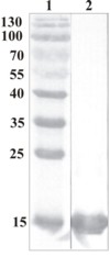
Figure 1. Western Blot testing of anti-CDNF polyclonal antibody. Line 1. PageRuler Prestained Protein Ladder (#SM0671 Fermentas); Line 2. Recombinant CDNF expressed into the supernatant of CHO cell culture medium.
A
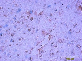 B
B
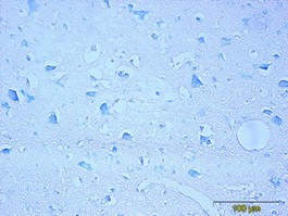
 B
B
Catalogue #
300-100
Name
Rabbit polyclonal antibody to human CDNF
Target
Human CDNF
Target Description
Recombinant human CDNF protein produced using CHO-based Icosagen Cell factory Ltd. proprietary suspension cell line. Purified from cell culture supernatant.
Alternative Names
ARMETL1
Uniprot ID
Q49AH0
Clonality
Polyclonal
Class
IgG
Reactivity
Human, mouse; others not tested
Application
ELISA, WB, IF, IHC-P
Protocol
ELISA:
0,1-0,2 µg/ml
WB:
0,1-0,2 µg/ml
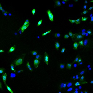
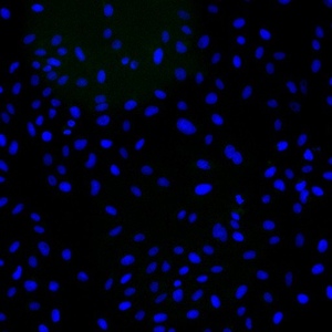
IF:
0,3-2 µg/ml
IHC-P:
5-10 µg/ml (on formalin fixed, paraffin-embedded tissues, antigen retrieval)
Purification
CDNF-affinity purification
Buffer
Ammonium sulphate, saturated (PBS pH 7.4)
Shipping
This product is shipped in non-frozen liquid form in ambient conditions
Storage
Store at +4°C upon receipt. As product is (NH 4)2SO4 precipitate, mix well by pipetting or vortexing prior use
Background
CDNF is a trophic factor for midbrain dopamine neurons in vivo. It prevents the 6-OHDA- (Lindholm et al. 20007; Voutilainen et al., 2011) and MPTP-induced degeneration (Airavaara et al., 2012) of dopamine neurons in rodent models of Parkinson’s disease. When administered after 6-OHDA or MPTP –lesioning it restores the dopaminergic function and prevents degeneration of dopamine neurons in substantia nigra pars compacta
This product is for research use only
TECHNICAL ASSISTANCE
Please refer any technical questions to
technical.support@icosagen.com


© 2024 Icosagen
![]()