Search from website
Search from website
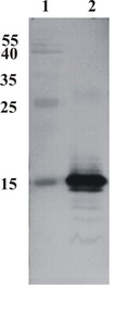
Figure 1. Western Blot testing of anti-CDNF monoclonal antibody (6G5). Line 1. PageRuler Prestained Protein Ladder (#SM0671 Fermentas). Line 2. Recombinant CDNF expressed into the supernatant of CHO cell culture medium.
A
 B
B
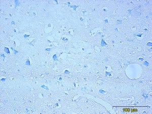
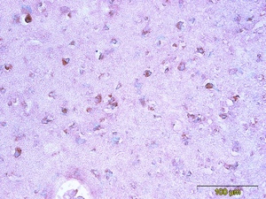
Figure 2. Immunohistochemistry testing of anti-CDNF monoclonal antibody 6G5. Analysis was performed using formalin-fixed paraffin-embedded human cerebral cortex tissue sections from Alzheimer’s disease patients. Tissue sections were boiled with sodium citrate buffer (pH 6) for antigen retrieval. Incubation with primary antibody at 5 µg/ml was performed overnight at 4°C. DAKO EnVisionTM Detection System, Peroxidase/DAB was used for visualization. Sections were counterstained with toluidine blue and mounted with Eukitt mounting medium. A. CDNF staining by monoclonal antibody 6G5; B. Negative staining without primary antibody.
A
 B
B
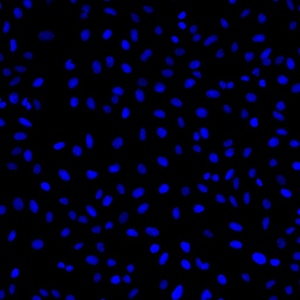
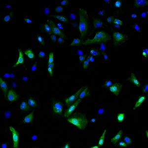
Figure 3. Immunofluorescence detection of human CDNF expressed in U2OS cells. CDNF was visualized using anti-CDNF antibody clone 6G5 at 1 µg/ml. Goat ant-mouse AlexaFluor488 was used as secondary antibody. For nuclear staining DAPI was used. ArrayScan VTI platform (Thermo Scientific) was used for image acquisition (10x objective). Composite picture was generated using pseudocolors green for CDNF specific signal and blue for nuclei. A. CDNF-expressing U2OS cells; B. Negative control (non-transfected U2OS cells).
Catalogue #
302-100
Name
Mouse mAb to human CDNF (clone 6G5)
Target
Human CDNF
Target Description
Recombinant human CDNF protein produced using CHO-based Icosagen Cell factory Ltd. proprietary suspension cell line. Purified from cell culture supernatant
Alternative Names
ARMETL1
Uniprot ID
Q49AH0
Clonality
Mouse monoclonal
Clone
6G5
Class
mIgG1
Reactivity
Human, no reactivity with mouse CDNF
Binds to the C-terminal part of the human CDNF (aa 126-187)
Application
ELISA, WB, IF, IHC
Protocol
ELISA:
0,06-0,2 µg/ml
WB:
0,25-1 µg/ml
IF:
0,3-10 µg/ml
IHC:
5-10 µg/ml (on formalin fixed, paraffin-embedded tissues)
Purification
Protein G purified
Buffer
PBS pH 7.4, with 0.1% sodium azide
Shipping
This product is shipped in non-frozen liquid form in ambient conditions
Storage
Store at – 20 or -70 °C upon receipt. Divide antibody into aliquots prior usage. Avoid multiple freeze-thaw cycles as product degradation may result.
Background
CDNF is a trophic factor for midbrain dopamine neurons in vivo. It prevents the 6-OHDA- (Lindholm et al. 20007; Voutilainen et al., 2011) and MPTP-induced degeneration (Airavaara et al., 2012) of dopamine neurons in rodent models of Parkinson’s disease. When administered after 6-OHDA or MPTP –lesioning it restores the dopaminergic function and prevents degeneration of dopamine neurons in substantia nigra pars compacta
This product is for research use only
TECHNICAL ASSISTANCE
Please refer any technical questions to
technical.support@icosagen.com


© 2024 Icosagen
![]()