Search from website
Search from website
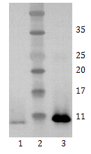
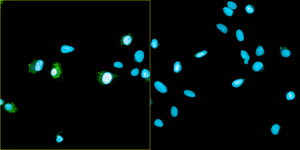
Figure 2. Immunofluorescence detection of hBDNF expression in U2OS cells by anti-BDNF monoclonal antibody 4C8. Antibody concentration 20 μg/ml. Goat anti-mouse AlexaFluor488 was used as secondary antibody. For nuclear staining DAPI was used. ArrayScan VTI platform (Thermo Scientific) was used for image acquisition (10x objective). Composite picture was generated using pseudocolors green for BDNF specific signal and blue for nuclei. A. proBDNF-expressing U2OS cells; B. Negative control (non-transfected U2OS cells).
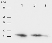
Figure 3. Western Blot testing of anti-BDNF monoclonal antibody 4C8. Antibody concentrations of 1 µg/ml was used. 2 Lanes 1 recombiant BDNF 1 µg , Lines 2 and 3 – neuron lysates. Photo courtesy of Indrek Koppel and Tõnis Timmusk, Tallinn Technical University, Institute of Gene Technology.
Catalogue #
328-100
Name
Mouse mAb to hBDNF (clone 4C8)
Target
Human BDNF
Target Description
Recombinant human BDNF protein purified from E. coli
Alternative Names
Abrineurin
Uniprot ID
P23560
Clonality
Mouse monoclonal
Clone
4C8
Class
mIgG1
Reactivity
Human, mouse, rat. guinea pig
Application
ELISA, WB, IF
ELISA:
0,05-0,1 µg/ml
WB:
0,2-1 µg/ml
IF:
20 µg/ml (weak signal)
Purification
Protein G purification
Buffer
PBS with 0.1% sodium azide
Shipping
This product is shipped in non-frozen liquid form in ambient conditions
Storage
Store at -20… -70 °C upon receipt. Divide antibody into aliquots prior usage. Avoid multiple freeze-thaw cycles.
Background
Brain-derived neurotrophic factor (BDNF) plays an important role in activity-dependent synaptic plasticity such as long-term potentiation. BDNF acts on certain neurons of the central nervous system and the peripheral nervous system, helping to support the survival of existing neurons, and encourage the growth and differentiation of new neurons and synapses
This product is for research use only
TECHNICAL ASSISTANCE
Please refer any technical questions to
technical.support@icosagen.com


© 2024 Icosagen
![]()